
Presentation
60-year-old male presents with hematuria and perineal/rectal pain.
Patient Data


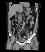
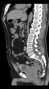
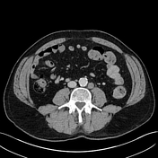
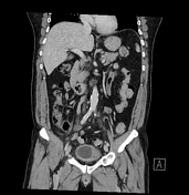
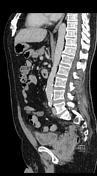
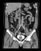
A large heterogenous fluid and soft tissue attenuating mass lies in the rectovesical pouch and is inseparable both from the distal colon and the prostate. There is extensive associated presacral, bilateral common and external iliac and left para-aortic lymphadenopathy which is likely malignant.
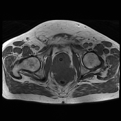
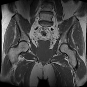
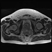
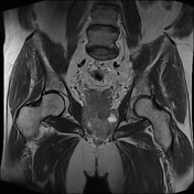
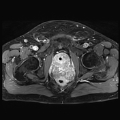
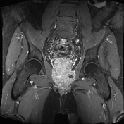

Large heterogenous partially necrotic mass within the pelvis measuring 75 x 62 x 113mm, favored arising from the prostate, involving the pelvic floor, inseparable from the prostate and seminal vesicles with posterior displacement of the rectum. There is a loss of the fat plane between the lesion and the anterior aspects of the left internal obturator and levator muscles as well as the rectum extending from 10 o'clock to 3 o'clock position. This lesion appears continuous with the base of the bladder.
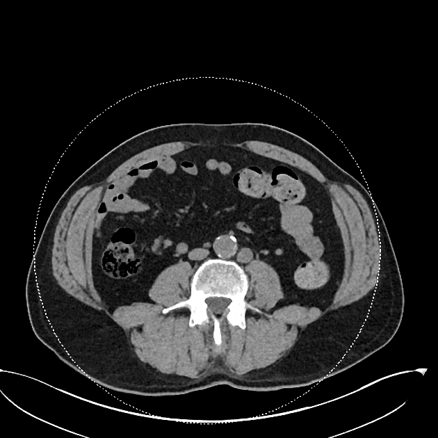
Bulky tumor deposits along the mesorectal vessels with bilateral external, internal and mesorectal lymphadenopathy. No inguinal lymphadenopathy. Malignant appearing left para-aortic lymph node.
Conclusion:
- appearance highly suspicious for aggressive prostate malignancy with an invasion of the bladder base the lower rectum and possibly the pelvic sidewall
- tumor deposits with vascular invasion throughout the mesorectum with bilateral malignant iliac and left para-aortic lymphadenopathy
- lesion amenable to trans-rectal biopsy
Case Discussion
This gentleman underwent a trans-rectal prostate biopsy. One out of six cores showed smooth muscle extensively infiltrated by small cell neuroendocrine carcinoma.
Small cell carcinoma of the prostate accounts for <1% of all cases of prostate cancer. Serum PSA levels are typically low. Median survival is 1-2 years from the time of diagnosis. Treatment involves a combination of chemo- and radiotherapy. Hormone therapy does not tend to work well in these patients. Most small cell prostate cancers have spread outside the prostate gland when they are diagnosed, so surgery is often not possible.


 Unable to process the form. Check for errors and try again.