
- Note: This case has been tagged as "legacy" as it no longer meets image preparation and/or other case publication guidelines.
From the case:
von Hippel Lindau


Download
Info
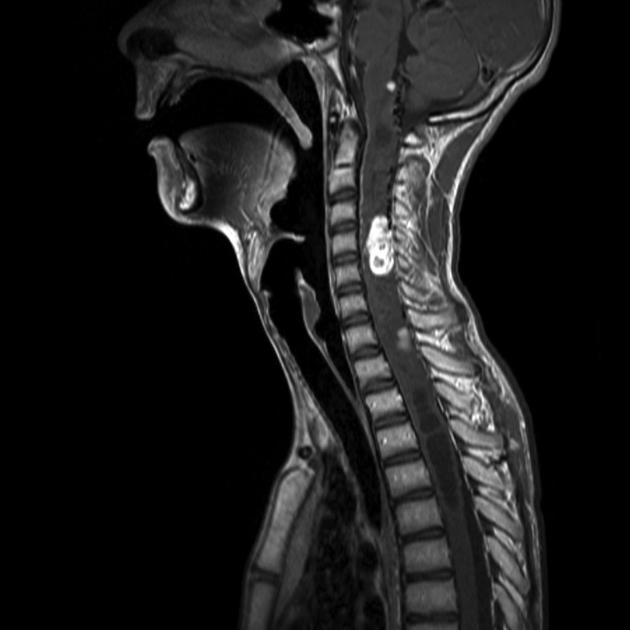
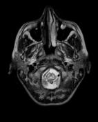
Selected MRI images demonstrate multiple enhancing intramedullary lesions, the largest at C4/5, with an associated syrinx.
From the case:
von Hippel Lindau

Download
Info
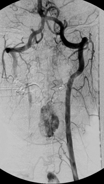
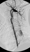
Catheter angiography demonstrates the cord lesions to be very hypervascular consistent with hemangioblastoma.
From the case:
von Hippel Lindau


Download
Info
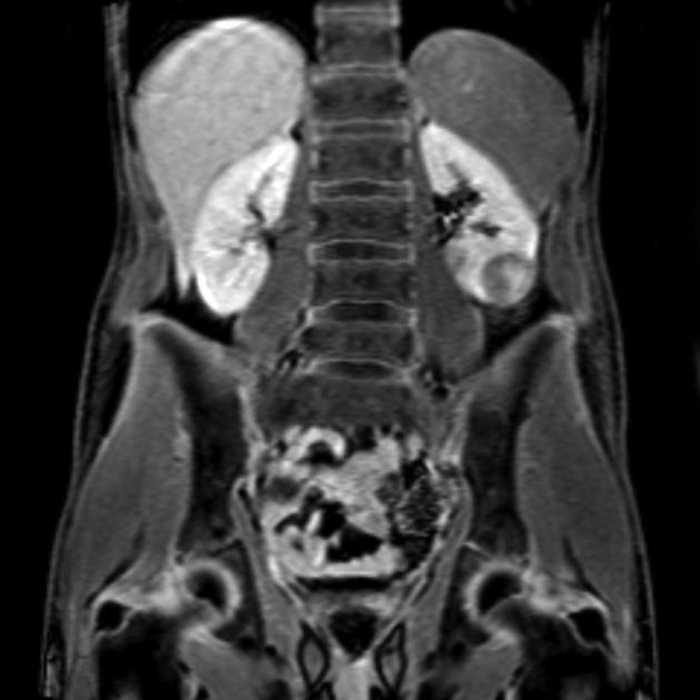
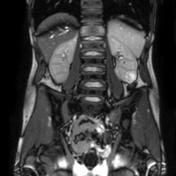
MRI demonstrates a left inferior pole renal mass consistent with a renal cell carcinoma.
Case Discussion
This individual has established von Hippel Lindau disease with a renal cell cancer on the left and cervical cord hemangioblastomas with an associated syrinx.


 Unable to process the form. Check for errors and try again.