
Presentation
Respiratory wheeze.
Patient Data
Age: 1 year
Gender: Male
From the case:
Thoracic teratoma - infant


Download
Info
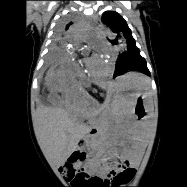
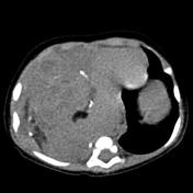
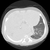
There is a large heterogeneous lesion with fat and calcific elements occupying right subphrenic space with superior extension into the thoracic cavity causing marked pressure effect on the right lung and left-sided shifting of cardiomediastinal structures and inferior extension compressing the liver and right kidney. Associated right-sided pleural effusion and the passive collapse of the right lung lower lobe also are noted.
Case Discussion
Selected axial and coronal chest CT confirms the presence of a huge hypodense well defined right-sided thoracoabdominal mass, with calcification and fat within it, the appearance is highly suggestive of teratoma.


 Unable to process the form. Check for errors and try again.