
From the case:
Callosal dysgenesis and lipoma
Download
Info
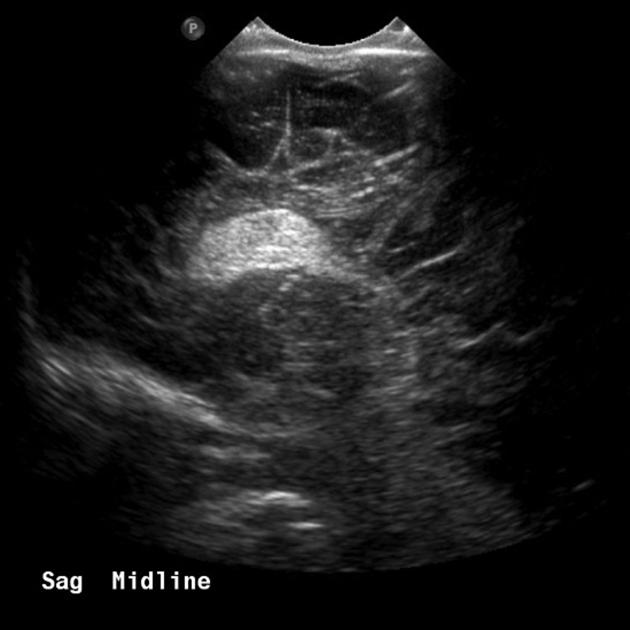
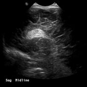
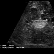
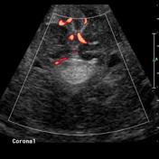
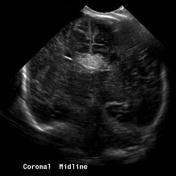
Transcranial ultrasound of an infant demonstrates a hyperechoic mass in the midline associated with callosal dysgenesis.
From the case:
Callosal dysgenesis and lipoma

Download
Info

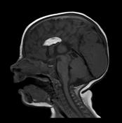
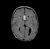
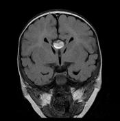
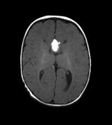
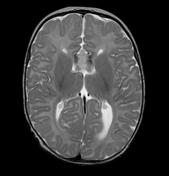
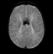
MRI demonstrates dysgenesis of the corpus callosum, with only the genu seen and without a formed rostrum. In the midline is an interhemispheric lesion which is of high T1 signal, and demonstrates signal drop out on fat-saturated sequences consistent with a pericallosal lipoma.
Case Discussion
Typical features of corpus callosum dysgenesis with an associated tubulonodular pericallosal lipoma.


 Unable to process the form. Check for errors and try again.