
Presentation
Right iliac fossa pain.
Patient Data


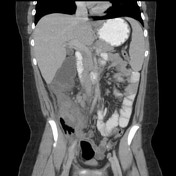
Large fluid collection located in the right lower quadrant with air-fluid levels, extending to the subhepatic space surrounded by thick irregular enhancing wall and surrounding fat stranding. No phlegmon.
Dilated fluid-filled appendix with a small calcified appendicolith at the base of the collection.
Bowel wall thickening in the right lower quadrant, with mild dilatation of the small bowel proximally.
Multiple enlarged mesenteric lymph nodes are seen the largest measure two centimeters in short axis.
Mild splenomegaly. Normal pancreas and both kidneys. Underfilled urinary bladder.
Case Discussion
This young patient presenting through the emergency room with right lower quadrant pain was suspected to have acute appendicitis. CT of the abdomen and pelvis with IV contrast was requested demonstrated dilated appendix with appendicolith and fluid collection in the appendicular region with air-fluid levels. Findings of appendicitis with appendicular abscesses.


 Unable to process the form. Check for errors and try again.