
Presentation
Abnormal chest radiograph done for ? chest infection. Chest CT was requested.
Patient Data
Age: 5 months
Gender: Female
From the case:
Polysplenia syndrome - Bochdalek hernia


Download
Info
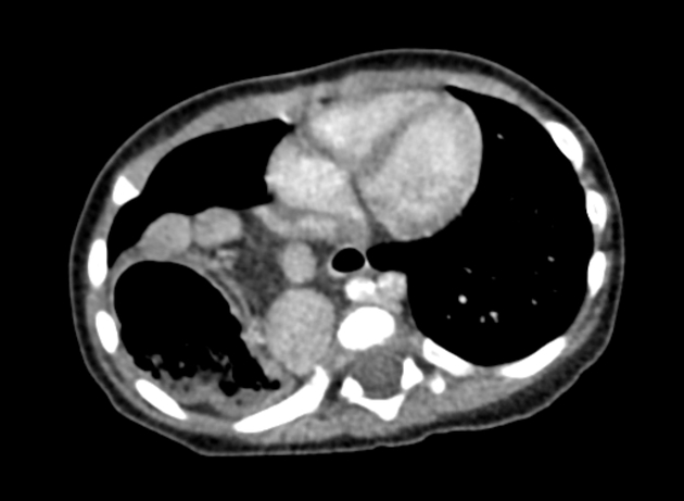
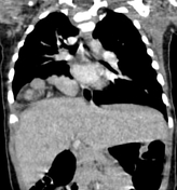
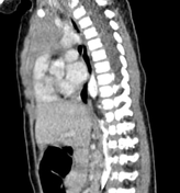
The CT scan demonstrates:
- right posterior diaphragmatic defect with intrathoracic herniation of:
- the stomach which is located on the right
- and numerous well-defined nodules of various size and homogeneous density (multiple splenules)
- no parent spleen is seen in the left hypochondrium
- intrahepatic IVC interruption with azygos/hemiazygos continuation
- midline/transverse liver
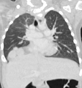
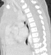
- bilateral bilobed lungs on lung window
- bilateral hyparterial bronchi
- preduodenal portal vein
- the body and tail of the pancreas are difficult to identify from the bowel loops that have the same density
- no evidence of midgut malrotation
Case Discussion
CT features of a polysplenia syndrome with associated Bochdalek hernia. This association is considered uncommon and extremely rare 1.
The patient was referred to a cardiopaediatrician to rule-out an associated congenital heart disease.
Additional contributor: ZE. Boudiaf, MD, CHU, Constantine, Algeria


 Unable to process the form. Check for errors and try again.