
Presentation
Abdominal pain and distension.
Patient Data
Age: 65 years
Gender: Male
From the case:
Pseudomyxoma peritonei

Download
Info

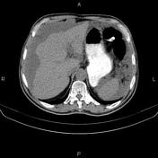
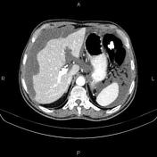
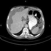
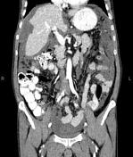
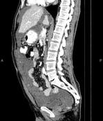
There are multiple low-attenuating mass like lesions with loculated fluid throughout the peritoneum, omentum and mesentery, accompanied by scalloping of visceral surfaces, particularly the liver. No calcification is present.
A few simple cortical cysts are seen at both kidneys, less than 18mm.
The prostate gland is enlarged.
Degenerative changes as osteophytosis are seen at the lumbar spine.
Case Discussion
Features on CT scan are consistent with diffuse peritoneal tumoral seeding most likely pseudomyxoma peritonei. It is most commonly caused by a mucinous tumor of the appendix1 .


 Unable to process the form. Check for errors and try again.