
Presentation
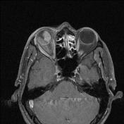
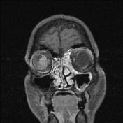
Right sided vision loss. Mass on ophthalmological examination.
Patient Data
Age: 60 years
Gender: Male
Note: This case has been tagged as "legacy" as it no longer meets image preparation and/or other case publication guidelines.
From the case:
Choroidal melanoma


Download
Info
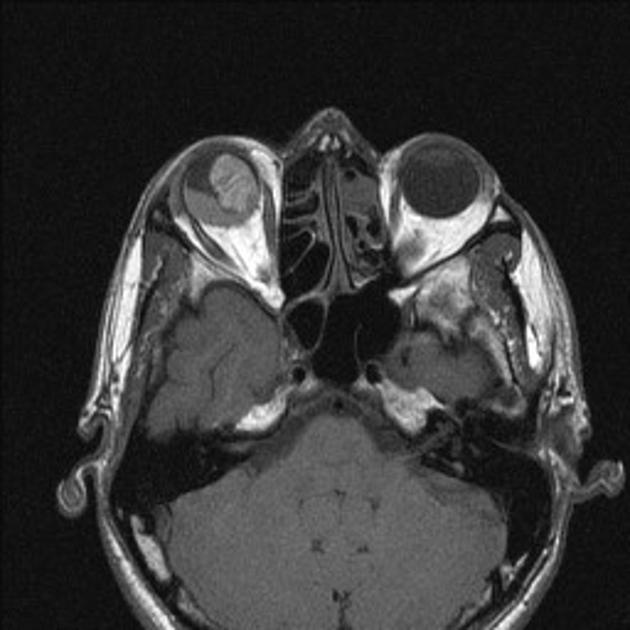
Contrast enhanced MR image shows a subretinal enhancing mass lesion with retinal detachment, with typical T1 hyperintense signal.
Case Discussion
Patient was operated subsequently, and histopathological diagnosis of choroidal melanoma was confirmed.


 Unable to process the form. Check for errors and try again.