
Presentation
MAG-3 scan performed for poor renal function (stage 4 CKD) and photopenic region of the left kidney leads to a CT. Known type 2 diabetes mellitus.
Patient Data
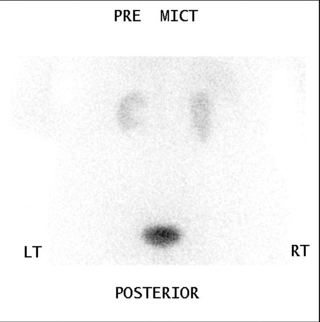
A single, premicturition, image of a complete MAG-3 renogram has been provided to demonstrate the potentially concerning finding of a focal photopenic area in the pelvis of the left kidney, which is otherwise normal in contour. The right kidney appears to be malrotated.


Oral contrast medium, no IV contrast medium due to marked renovascular disease.
Unremarkable appearances of the unenhanced liver, gallbladder, spleen, pancreas and adrenals.
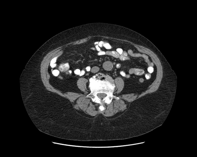
6 cm interpolar renal cyst on the right. No hydronephrosis. On the left the kidney is irregular in outline - this may be related to scarring or lobulation but no mass lesion.
Small right of midline supraumbilical ventral hernia containing omental fat only.
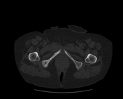
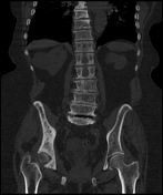
Diffusely expanded bone of the right hemipelvis with thickened cortices and heterogeneous bone texture likely representing Paget disease.
Conclusion
In view of the Pagetoid bone, bone scintigraphy was performed.
No concerning renal findings.
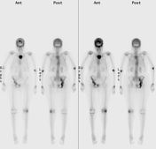
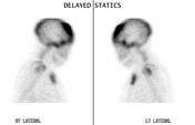
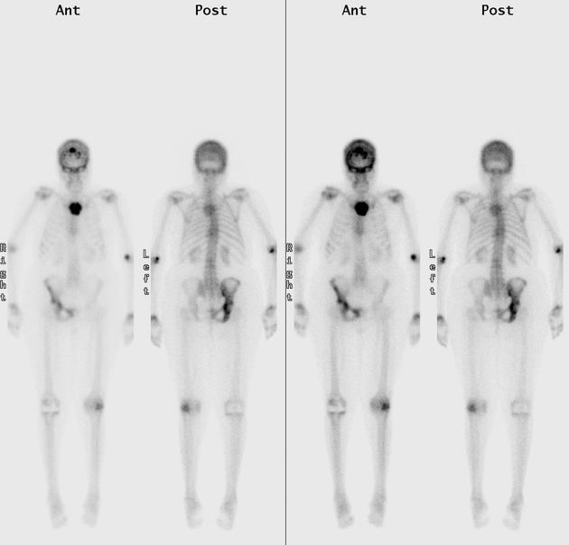
Increased activity of the skull vault, especially frontal bones, and the anterior skull base. Also increased activity of the superior sternum (likely manubrium, not body). Diffusely increased activity of the right hemipelvis.
Other findings
- photopenia of the right knee due to a TKR
- degenerative joint disease
- photopenic focus of the left kidney again seen
Conclusion:
Bony appearances likely related to Paget disease, however radiographic correlation was performed for the skull, sternum and pelvis (despite CT performed only two months' earlier).
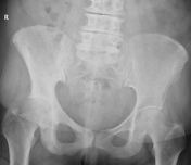
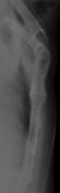

Typical Pagetoid appearances of the skull, manubrium sterni and pelvis.
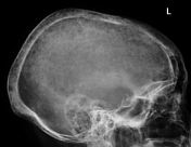
- skull
- osteoporosis circumscripta cranii
- diploic widening
- cotton wool appearance
- pelvis
- right-sided
- involvement of ischium, ilium and pubis
- Kohler teardrop is not seen
- pelvic brim sign
Case Discussion
Paget's disease remains common in older patients with European ethnicity affecting 3-4% over 40 year olds. It is rare in those from Scandinavia or with Asian/African ethnicities 1,2.
Pagetoid involvement of the sternum is rare and when it occurs can make procedures necessitating a sternotomy, e.g. CABG, challenging 2.
An interesting learning point demonstrated is the poor sensitivity of bone scintigraphy for Paget disease. If one compares the bone scan with the radiographs and CT, several parts of the right hemipelvis show little activity on the functional study, whilst the structural imaging shows diffuse involvement of all three bones of the hemipelvis. Research has shown that scintigraphy tends to show a lack of tracer uptake in quiescent areas of the disease 1.
This case is a nice example of how concern about one thing, i.e. renal impairment, leads to a test MAG-3 which raises concerns of a renal lesion, leading to a CT, which in turn raises concerns about the skeleton leading to further imaging.


 Unable to process the form. Check for errors and try again.