
Presentation
Incidental gastric mass seen on surveillance vascular imaging.
Patient Data
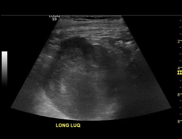
Longitudinal US of the Lt Upper Quadrant
Heterogeneously hypoechoic mass in the left upper quadrant, the location of which is difficult to ascertain on this cine sweep due to lack of anatomic landmarks.


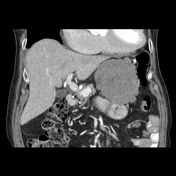
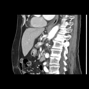
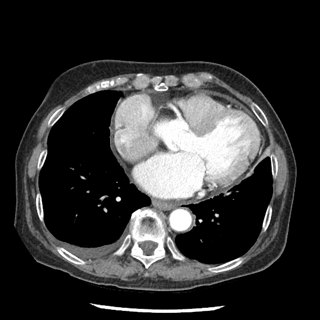
Contrast-Enhanced CT of the Abdomen
Heterogeneous, enhancing lobulated mass arising from the wall of the greater curvature of the stomach and extending into the gastrohepatic space.
Scattered, ill-defined hypovascular lesions in the lateral segments of the left hepatic lobe and subcentimeter hypervascular lesions in hepatic segments 2 and 4b.
Trace right pleural effusion.
Case Discussion
An endoscopic ultrasound-guided fine needle aspiration of the gastric mass showed low-grade spindle neoplasm, consistent with gastrointestinal stromal tumor (GIST).
The patient was treated with imatinib and subsequently surgical resection with sleeve gastrectomy. Surgical pathology showed a 6.9 cm CD117+ and DOG-1+ GIST with negative margins.


 Unable to process the form. Check for errors and try again.