
Presentation
Abdominal pain and dyspepsia.
Patient Data


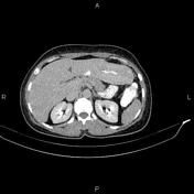
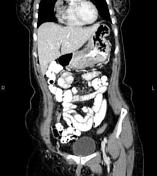
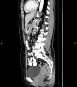
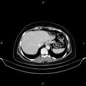
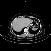
Wall thickening and enhancement are seen at the gastric body and antrum. Perigastric fat stranding and small adenophaty are also noted.
Mild degenerative changes as osteophytosis are seen at the lumbar spine.
Grade I spondylolisthesis of L5 on S1 is present with bilateral spondylolysis.

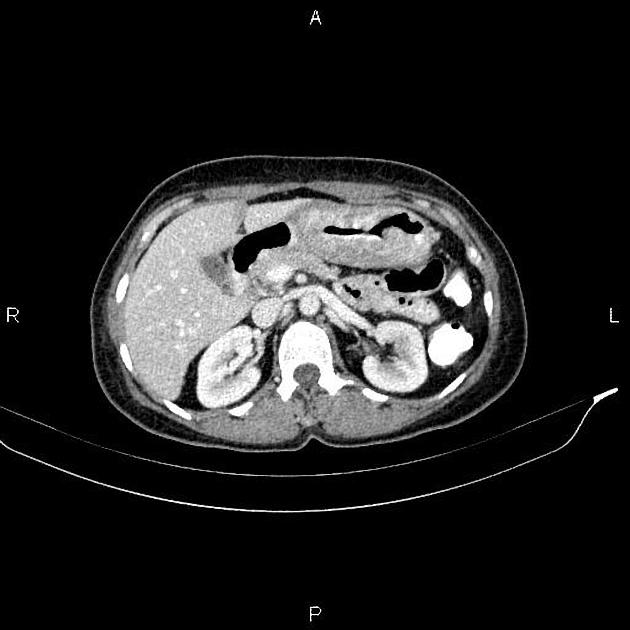
Post surgical control CT after 15 months
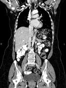
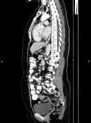
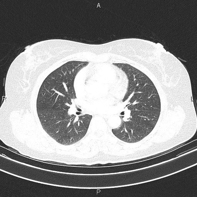
Post-operative changes are seen due to total gastrectomy and esophagojejunostomy. There are no sign of local tumoral recurrence at anastomotic site and no regional lymphadenopathy.
Mild degenerative changes as osteophytosis are seen at the lumbar spine.
Grade I spondylolisthesis of L5 on S1 is present with bilateral spondylolysis.
Case Discussion
Pathology proven and operated case of gastric adenocarcinoma. CT is currently the staging modality of choice because it can help identify the primary tumor, assess for the local spread, and detect nodal involvement and distant metastases.


 Unable to process the form. Check for errors and try again.