
Presentation
Abdominal pain and diarrhea.
Patient Data
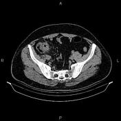


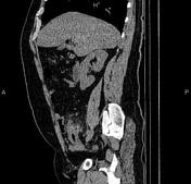

The hepatic attenuation value is less than of the spleen (35HU vs. 41HU on non-contrast images), suggesting fatty liver.
Terminal ileum shows inflammatory change and its wall thickness increased up to 11 mm. Stranding was seen in adjacent fat. Also, an enlarged lymph node measuring 27×19 mm was noted in this region. Appendix appears normal.
Case Discussion
Terminal ileitis in a 45 years patient with abdominal pain and diarrhea. The patient underwent colonoscopy evaluation and tissue exam confirms Crohn disease.
CT scan is commonly the first imaging assessment of those patients in the setting of acute abdomen, or it can be also applied to the reassessment of complications in patients with known Crohn disease.


 Unable to process the form. Check for errors and try again.