
Presentation
Abdominal pain and progressive distension.
Patient Data
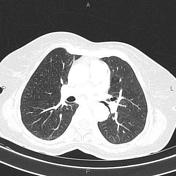


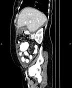

A few atelectatic bands are seen at both lung particularly at bases.
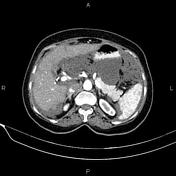
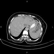
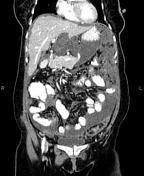
Post-operative changes are seen due to right hemicolectomy.
Moderate ascites is present. There are multiple low attenuating lesions causing scalloping the liver margin. Omental thickening and numerous varying sized omental soft tissue masses are also evident. Additionally, a 32×20 mm cystic solid lesion is observed at right inguinal region.
Degenerative changes as osteophytosis are seen at the thoracolumbar lumbar spine.
Case Discussion
Evidence of right hemicolectomy (pathology proven mucinous adenocarcinoma of colon) and pseudomyxoma peritonei that refers to syndrome of progressive intraperitoneal accumulation of mucinous ascites related to a mucin-producing neoplasm.
On CT scan evaluation pseudomyxoma peritonei is characterized by loculated collections of fluid which accumulate along peritoneal surfaces, classically resulting in a scalloped appearance of coated abdominal organs and omental caking.


 Unable to process the form. Check for errors and try again.