
Presentation
Abdominal pain and dyspepsia.
Patient Data


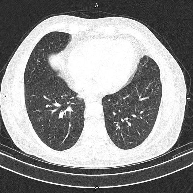
A few small nodules are seen at both lungs less than 4 mm.
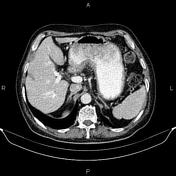
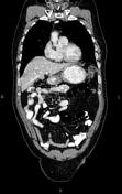
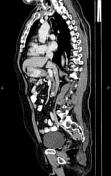
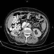
Increased wall thickness suggestive of tumoral infiltration is present in the lesser curvature of the stomach. Mild perigastric fat stranding and small volume regional lymphadenopathy are evident.
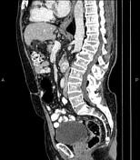
The prostate gland is enlarged.
Degenerative changes as osteophytosis are seen at the thoracolumbar spine.
Partially collapse is noted at T12 and L1 vertebrae.

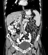
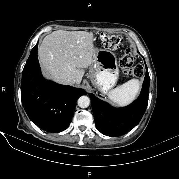
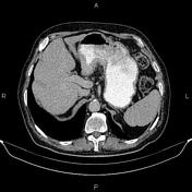
Post-operative change is seen as distal partial gastrectomy and gastrojejunostomy. There is no sign of local tumoral recurrence at the surgical site.
The prostate gland is enlarged.
Degenerative changes as osteophytosis are seen at the thoracolumbar spine.
Partially collapse is noted at T12 and L1 vertebrae.
Case Discussion
Pathology proven gastric lesser curvature adenocarcinoma. The patient underwent a distal partial gastrectomy and gastrojejunostomy and chemotherapy.
CT is currently the staging modality of choice because it can help identify the primary tumor, assess for the local spread, and detect nodal involvement and distant metastases.


 Unable to process the form. Check for errors and try again.