Presentation
Left flank pain and hematuria
Patient Data
Age: 40 years
Gender: Female
From the case:
Renal cell carcinoma
![](https://prod-images-static.radiopaedia.org/images/54332129/77ded80cf1503deb3c38b6ee1f74db71d4c5d06d5f65ea0217fd221226f00cea_thumb.jpeg)
![This study is a stack](/packs/stack-YQKLCKBI.gif)
![](https://prod-images-static.radiopaedia.org/images/54332164/28b60c27afbd709eb00d5760048f10bc0260e538e334e1b59d560dd4997b69c7_thumb.jpeg)
![This study is a stack](/packs/stack-YQKLCKBI.gif)
![](https://prod-images-static.radiopaedia.org/images/54332202/ae234e4cceb0e7754e42cbbf7c4c900d51fd78d1032a7f56e5e115fbd6c87ba4_thumb.jpeg)
![This study is a stack](/packs/stack-YQKLCKBI.gif)
![](https://prod-images-static.radiopaedia.org/images/54332242/6d0fe284f28d2936656ffb87a5fa135ec1427868790dc1115f13349cb5463f6a_thumb.jpeg)
![This study is a stack](/packs/stack-YQKLCKBI.gif)
![](https://prod-images-static.radiopaedia.org/images/54332282/35ef8620f6f35f8a7e5eb94b4d8a8e03c69e5272ee8af917ee300b5580e7d961_thumb.jpeg)
![This study is a stack](/packs/stack-YQKLCKBI.gif)
![](https://prod-images-static.radiopaedia.org/images/54332322/105059170db341c331ecaf90bf7ac130b507813437131c2f5c74261ecab2d5bd_thumb.jpeg)
![This study is a stack](/packs/stack-YQKLCKBI.gif)
![](https://prod-images-static.radiopaedia.org/images/54332357/0a5ee490497552f29cb38317b7e7d5af427ea1684cab7fdb817a65ac5847559e_thumb.jpeg)
![This study is a stack](/packs/stack-YQKLCKBI.gif)
![](https://prod-images-static.radiopaedia.org/images/54332388/b6b7175e71e6f3215345087cd904f3ac99899b90145f38e93ede19c7fe807758_thumb.jpeg)
![This study is a stack](/packs/stack-YQKLCKBI.gif)
![](https://prod-images-static.radiopaedia.org/images/54332438/ea1ebe047de307445a39cb777c692ad7d140f6e2c268ced871ebc42169469ad6_thumb.jpeg)
![This study is a stack](/packs/stack-YQKLCKBI.gif)
![](https://prod-images-static.radiopaedia.org/images/54332504/b6ef0d19ec477890d29d1bd36e513313d8a21fd59ad12ede18bef9e1e74c1e7e_thumb.jpeg)
![This study is a stack](/packs/stack-YQKLCKBI.gif)
![](https://prod-images-static.radiopaedia.org/images/54332568/734f98b9839bd4a73abf2bb26df0d887250a1c2c4cb6a3473f2372b987dd66a7_thumb.jpeg)
![This study is a stack](/packs/stack-YQKLCKBI.gif)
![](https://prod-images-static.radiopaedia.org/images/54332616/db7253bd0432701028edba77a6ce985505c3edc1a208bf15994d778da6642d06_thumb.jpeg)
![This study is a stack](/packs/stack-YQKLCKBI.gif)
![](https://prod-images-static.radiopaedia.org/images/54332702/5e2350906811c2a6d2ae7f82d62b0a958169ec4f5ccdc7c9fc906378fefc13a9_thumb.jpeg)
![This study is a stack](/packs/stack-YQKLCKBI.gif)
![](https://prod-images-static.radiopaedia.org/images/54332764/07423905b4cd2a14c83af3b145d1888f7e9238e7279b868753abf05a1e4df708_thumb.jpeg)
![This study is a stack](/packs/stack-YQKLCKBI.gif)
Download
Info
![](https://prod-images-static.radiopaedia.org/images/54332129/77ded80cf1503deb3c38b6ee1f74db71d4c5d06d5f65ea0217fd221226f00cea_big_gallery.jpeg)
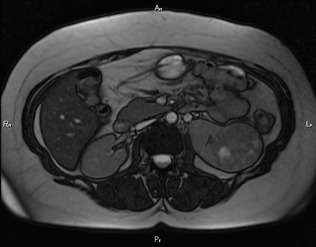
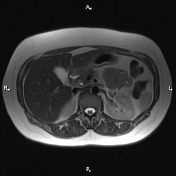
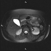
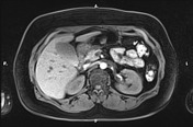
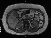
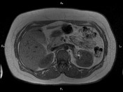
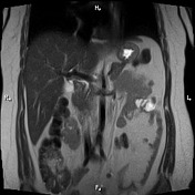
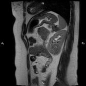
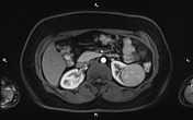
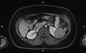
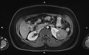
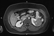
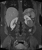
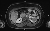
A well defined partially exophytic heterogeneous solid mass is seen at mid to lower pole of left kidney. After contrast media injection, it shows mild heterogeneous enhancement with some necrotic components within. There is no fat stranding in perirenal space and extension to left renal vessels is not seen.
Case Discussion
The patient underwent left nephrectomy and histopathology confirms renal cell carcinoma which is the most common malignant renal tumor.