
Presentation
Abdominal pain and dyspepsia.
Patient Data
Age: 50 years
Gender: Male
From the case:
Gastric adenocarcinoma


Download
Info
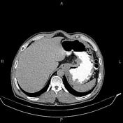
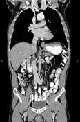
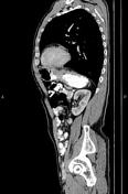
Increased wall thickness due to tumoral infiltration is present at gastric cardia, sub cardia and proximal of lesser curvature; accompanied by mild adjacent fat stranding and several small regional lymphadenopathies.
A 25 mm simple cortical cyst is noted at right kidney.
The prostate gland is enlarged.
Case Discussion
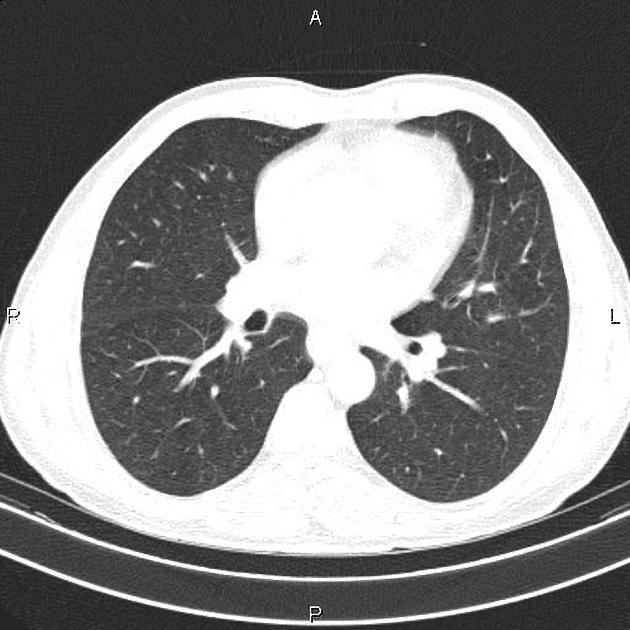
Pathology proven case of gastric adenocarcinoma with small regional lymphadenopathies. No local invasion; No detectable metastasis.
CT is currently the staging modality of choice because it can help identify the primary tumor, assess for the local spread, and detect nodal involvement and distant metastases.


 Unable to process the form. Check for errors and try again.