
Presentation
Abdominal pain and dyspepsia.
Patient Data
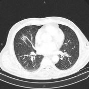


Few atelectatic bands are scattered at both lung fields.
Several small lymphadenopathies are seen at both hilar regions.
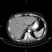
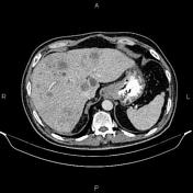
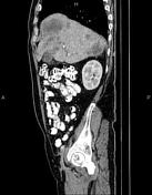
Increased wall thickness suggestive of tumoral infiltration is present at gastric cardia and sub cardia. Several lymphadenopathies are seen at peri gastric and porta hepatis regions. Additionally, multiple ill defined low enhancing masses are seen at liver.
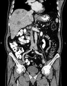
Few subcentimeter simple cortical cysts are seen at both kidneys.
The prostate gland is enlarged.
Degenerative changes as osteophytosis are seen at the thoracolumbar spine.
Case Discussion
Pathology proven case of gastric adenocarcinoma with peri gastric, porta hepatis and para hilar lymphadenopathies and hepatic metastasis.
CT is currently the staging modality of choice because it can help identify the primary tumor, assess for the local spread, and detect nodal involvement and distant metastases.


 Unable to process the form. Check for errors and try again.