
Presentation
Abdominal pain and dyspepsia.
Patient Data

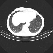

A few small calcified nodules less than 4mm scattered at both lung fields. There are also several atelectatic bands scattered bilaterally.
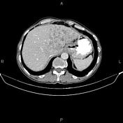
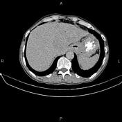
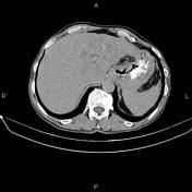
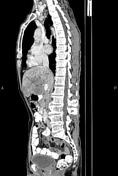
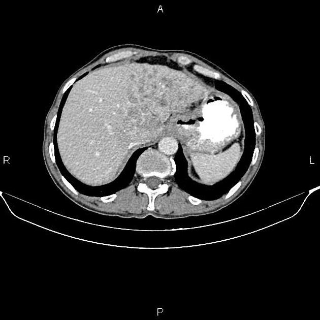
Increased wall thickness due to tumoral infiltration is present at esophagogastric junction, gastric cardia and sub cardia; accompanied by several regional lymphadenopathies with maximum SAD of 25 mm. Additionally, multiple low enhancing masses are seen at liver less than 60 mm.
Diffuse but mild urinary bladder wall thickening is present.
The prostate gland is enlarged.
Degenerative changes as osteophytosis are seen at the thoracolumbar spine.
Case Discussion
Pathology proven esophagogastric adenocarcinoma with regional lymphadenopathies and hepatic metastasis.
CT is currently the staging modality of choice because it can help identify the primary tumor, assess for the local spread, and detect nodal involvement and distant metastases.


 Unable to process the form. Check for errors and try again.