
Presentation
Abdominal pain and palpable mass on physical exam.
Patient Data


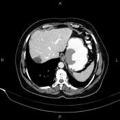
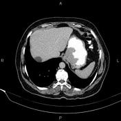
A 52 mm vegetative mass was seen at the gastric cardia.
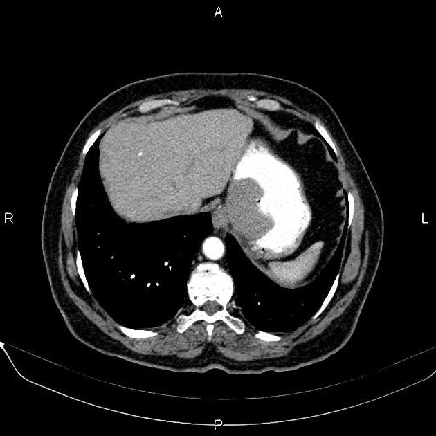
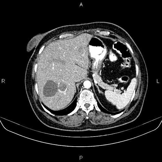
A 62 mm cyst was seen in the 7th segment of the liver.
Additionally, a hypodense lesion, 33 mm in diameter, was seen in the 7th segment of the liver. On the post contrast portal venous phase, a peripheral dense, spotty enhancement was noted in the mass. In the delayed phase the attenuated areas spread through the mass and show filling in. The density of the lesion is almost the same as that of aorta in all phases of dynamic CT.
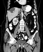
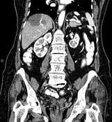
A 40 mm cyst was present in the mid-portion of right kidney.
Uterus and ovaries were not seen (prior surgery).

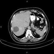
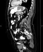
Post-operative change are seen due to proximal partial gastrectomy. There is no sign of tumoral remnant or recurrence at surgical site.
There is a 62 mm thin-walled cyst in the 7th hepatic segment.
Additionally, a 33 mm hypodense mass is noted in the 7th segment which shows early peripheral nodular enhancement with centripetal filling.
A few cortical cysts are seen in the kidneys.
The uterus and ovaries are not seen due to prior resection.
Case Discussion
Pathology proven case of gastric gastrointestinal stromal tumor (GIST) which is the most common mesenchymal tumor of the gastrointestinal tract.
Enhancement pattern of hepatic mass compatible with hemangioma. Simple hepatic cyst is also evident.


 Unable to process the form. Check for errors and try again.