
Presentation
Respiratory distress
Patient Data


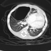
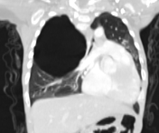
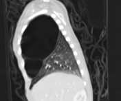
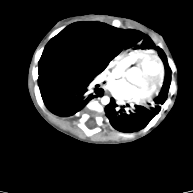
Pure multicystic mass lesion almost replacing right upper lobe and middle lobe. No visible communication with the bronchial system.
No soft tissue component or abnormal vasculature. It is causing a contralateral mediastinal shift.
The rest of the lungs are clear.
There is no evidence of pleural thickening or effusion. No pneumothorax.
Case Discussion
The large multicystic lesion is consistent with a type I congenital pulmonary airway malformation.
CT has a number of roles in the management of CPAMs. First, it more accurately delineates the location and extent of the lesion. Secondly, and most important in surgical candidates, CT angiography is able to identify systemic arterial supply if present.
This case is also contributed by Dr M.M.S.Hoshang.


 Unable to process the form. Check for errors and try again.