
Presentation
Chronic hepatitis B.
Patient Data


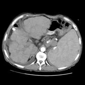

Triphasic liver CT was conducted and reveals the liver to have increase density with irregular nodular surface and parenchyma, consistent with cirrhosis.
No enhancing nodule seen during the arterial phase to suggest HCC.
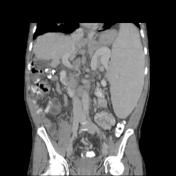
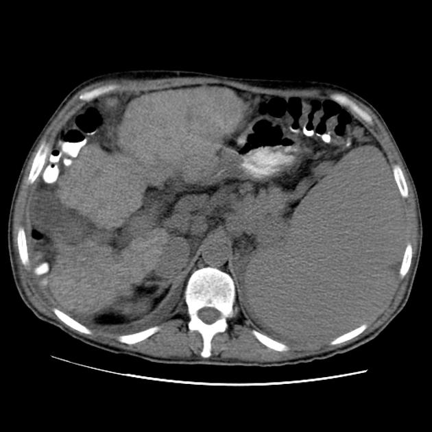
Huge splenomegaly is seen. Distended and engorged paraumbilical veins, which are seen radiating from the umbilicus across the abdomen to join the systemic veins (Caput Medusae) indicative of severe portal hypertension.
Multiple lienorenal and gastrosplenic varices.
Normally opacified portal, SMV and splenic veins. No evidence of cavernous transformation of the portal vein.
Moderate right sided pleural effusion.
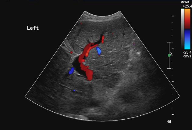
US abdomen showed the liver to have irregular surface with coarse nodular echotexture, suggestive of cirrhosis.
Patent portal vein.
Case Discussion
Known case of chronic hepatitis B infection with liver cirrhosis and signs of portal hypertension.


 Unable to process the form. Check for errors and try again.