
Presentation
Known melanoma. Metastases?
Patient Data
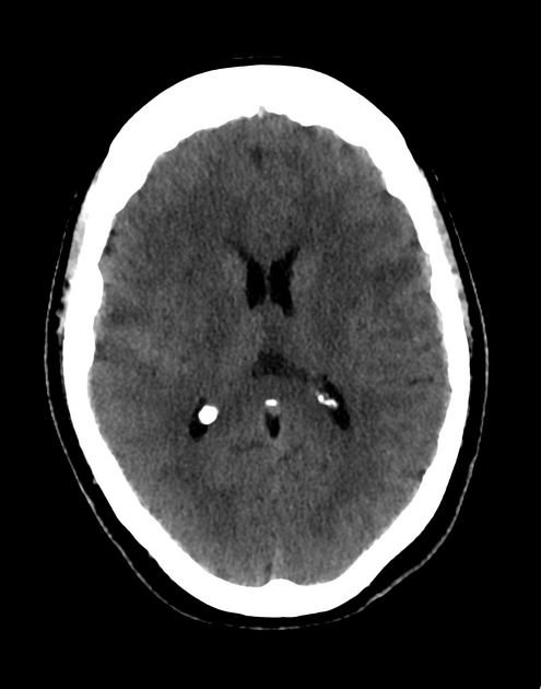
Right temporal and left frontal hyperdense metastatic deposits measuring 1.2 cm and 2.2 cm with perilesional edema. No significant mass effect.

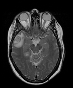
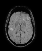
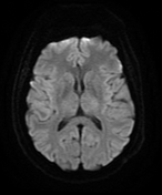
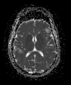

Widespread supra and infratentorial metastases, the largest 3.5 cm in the left parietal lobe with internal hemorrhage and layering of blood products. Additional right temporal lobe hemorrhagic metastasis as evidenced by severe susceptibility artifact.
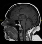
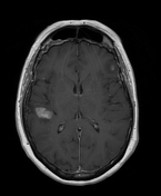
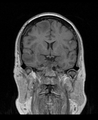
The smaller metastases are high signal on T1.
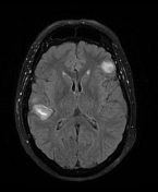
Moderate perilesional edema in relation to the larger metastases, but no significant mass effect.
Case Discussion
The MRI appearances of high T1 lesions are typical of metastatic melanoma due to the inherent high signal of melanin.
The high attenuation lesions on CT with corresponding intense susceptibility artifact on MRI indicate these are both in addition hemorrhagic metastases.
The appearances are typical of metastatic malignant melanoma consistent with the patient's medical history.


 Unable to process the form. Check for errors and try again.