
Presentation
Dysphagia and recurrent chest infection.
Patient Data

Chest x-ray is largely unremarkable. Minor patchy opacities in the left base are noted. A small gastric air bubble is visible.


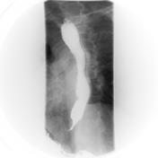
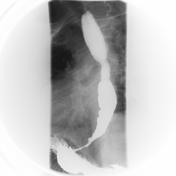
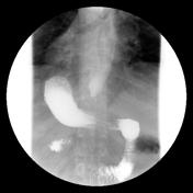
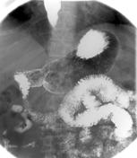
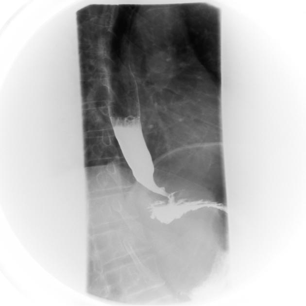
Upper GI study reveals uniform dilatation of the esophagus to the level of the gastro-esophageal junction, where fixed narrowing is noted (bird beak sign or rat-tail sign). Repeated observation by fluoroscopy confirmed failure of relaxation of the lower esophageal sphincter and prolonged retention of barium in the esophagus.

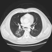
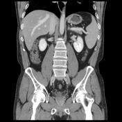
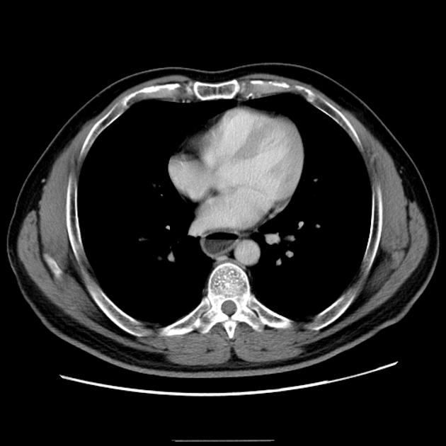
CT scan of the abdomen showed uniform dilatation of the esophagus with air-fluid level. Patchy consolidation in the upper segment of the right lower lobe likely due to aspiration.
Case Discussion
This case illustrates the typical features of achalasia, complicated by an aspiration pneumonitis, a relatively common complication.


 Unable to process the form. Check for errors and try again.