
Presentation
Chest pain, cough, and anorexia for 4 months.
Patient Data
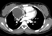


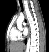

There is a well-defined, low attenuating lesion with wall discontinuous calcifications and internal fat loculi in the right side anterior mediastinum extending inferiorly to the level of the carina. The lesion is in close contact with the right atrium and is located anterolateral to the superior vena cava and anterior to the right superior pulmonary vein. The lesion measures 7 x 6 x 6.5 cm in size. No adiacent infiltrative changes. No enlarged mediastinal lymph nodes.
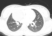
The normal aspect of the right and the left lung is noted showing normal volume. No evidence of air space consolidation nor nodular neither micronodular lesions in the pulmonary parenchyma.
Case Discussion
The imaging features are diagnostic of mediastinal teratoma.


 Unable to process the form. Check for errors and try again.