
Presentation
Abdominal pain and dyspepsia.
Patient Data


A few small lymphadenopathies are seen at both hilar regions, with SAD less than 14 mm. There are also several lymphadenopathies at subcarina and AP window with maximum SAD of 13 mm.
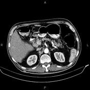
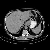
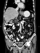
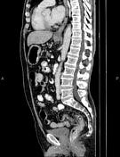
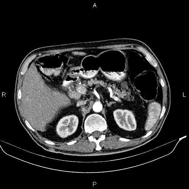
Increased wall thickness due to tumoral infiltration is present at gastric cardia, subcardia and proximal of lesser curvature; accompanied mild adjacent fat stranding and a few small regional lymphadenopathies with SAD less than 10 mm.
Multiple ill-defined low enhancing masses are seen at liver less than 50 mm.
A few thin walled non enhanced cysts are seen at pancreas less than 20 mm.
Several non enhanced simple cortical cysts are seen at both kidneys, with maximum diameters of 15 mm.
The prostate gland is enlarged.
Case Discussion
Gastric mass; pathology proven adenocarcinoma, with regional, mediastinal and hilar lymphadenopathies, diffuse hepatic metastases.


 Unable to process the form. Check for errors and try again.