
Presentation
History of shortness of breath.
Patient Data
Age: 25 years
Gender: Male


Download
Info
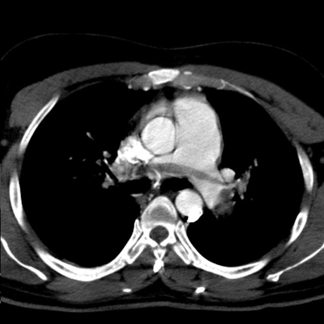
CTPA demonstrates saddle pulmonary embolism, extending to segmental and subsegmental branches of both pulmonary artery branches.
Dilated pulmonary artery and enlarged right ventricle, suggestive of secondary pulmonary arterial hypertension.
Download
Info
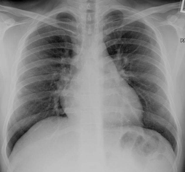
Bilateral peripheral pulmonary opacities.
Prominent right cardiac border.
Case Discussion
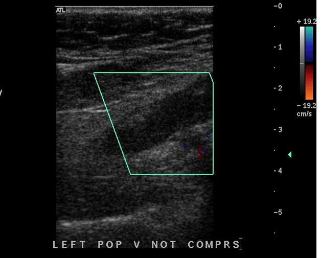
CTPA demonstrates saddle pulmonary embolism with secondary pulmonary arterial hypertension. DVT was confirmed by US Doppler study of the lower limb, as a possible etiology for pulmonary embolism.


 Unable to process the form. Check for errors and try again.