
Presentation
Low back pain.
Patient Data


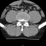
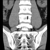
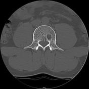
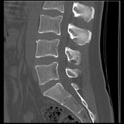
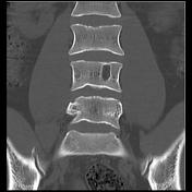
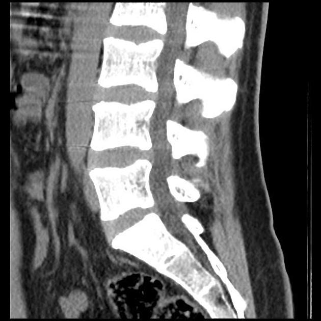
There is a well-defined intraosseous lesion of the L4 vertebral body of fatty density (mean density -62 HU) with sclerotic margin and small cortical interruption of the end-plate well-visualized on sagittal and coronal reformatted images. A small Schmorl node of the superior endplate of L3 is also noted.
The height of the vertebral bodies and disc spaces is preserved. No disc herniation is seen.
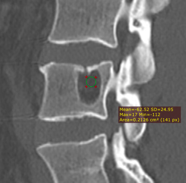
Annotated image showing the mean density of the intravertebral lesion (-62 HU).
Case Discussion
CT features of a well-defined intraosseous lesion of fatty density with a sclerotic peripheral rim of the L4 vertebral body with cortical interruption of the superior endplate suggestive of a giant fatty Schmorl node.
The main differential diagnosis is an intraosseous lipoma (usually the cortex of the vertebral endplates is intact).


 Unable to process the form. Check for errors and try again.