
Presentation
Shortness of breath and weight loss
Patient Data
Age: 75 years
Gender: Male
From the case:
Bronchioloalveolar carcinoma
Download
Info
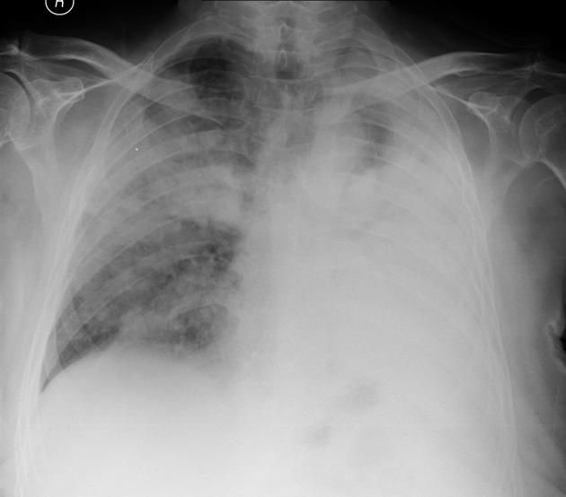
Widespread fluffy airspace opacities, with almost complete white-out of the mid and lower zones on the left.
From the case:
Bronchioloalveolar carcinoma

Download
Info
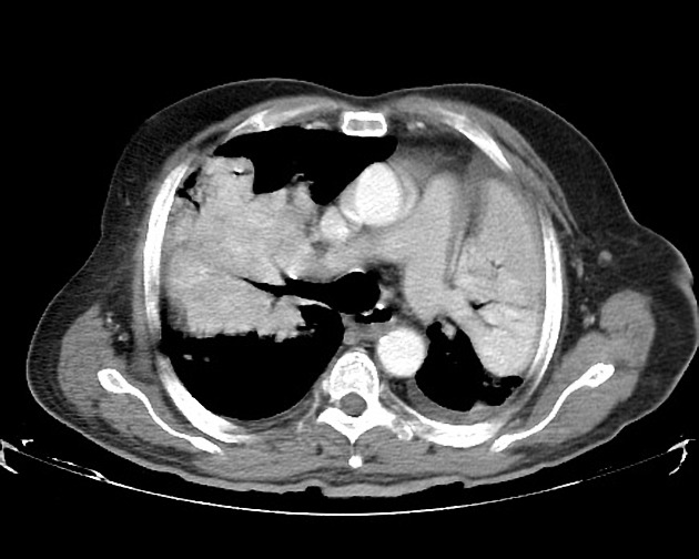
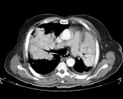
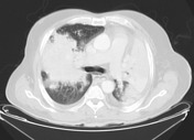
Extensive airspace opacities with numerous air bronchograms. Trace of pleural fluid on the left only. No significant nodal enlargement.
Please note terminology in this case (bronchoalveolar carcinoma) reflects practice at the time of publication and may not reflect current practice.
Case Discussion
Sputum, right and left main bronchus lavage were positive for malignant cells consistent with carcinoma. A Tru-cut biopsy was suggested by the pathologist to confirm the diagnosis of bronchoalveolar carcinoma, the patient's condition meant that this was not possible.


 Unable to process the form. Check for errors and try again.