
Presentation
Diffuse abdominal pain.
Patient Data


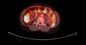
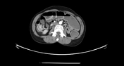
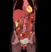
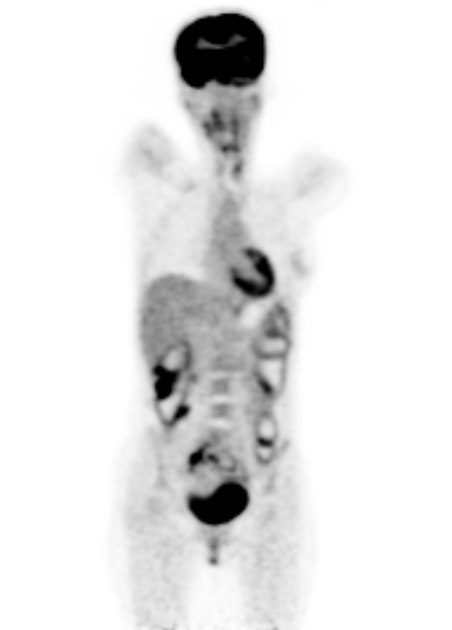
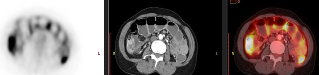
Multifocal increased FDG uptake by multifocal mild circumferential mural thickening involving small and large bowel loops and the rectum, most evident at the ascending colon and ileoceal junction.
Multiple FDG avid perirectal and internal iliac ymph nodes.
Multifocal increased FDG uptake by multifocal mild circumferential mural thickening involving small and large bowel loops and the rectum, most evident at the ascending colon and ileoceal junction.
Case Discussion
The case shows multifocal increased FDG uptake by multi-focal mild short segments of mural thickening involving the small and large bowel loops.
The patient underwent endoscopy and biopsy which revealed inflammatory bowel disease (Crohn disease).
PET-CT is useful in evaluating patients with inflammatory bowel disease, it helps to identify areas of active inflammation.


 Unable to process the form. Check for errors and try again.