
Presentation
History of left hemithyroidectomy for papillary thyroid cancer, now presents with a new right sided nodule.
Patient Data


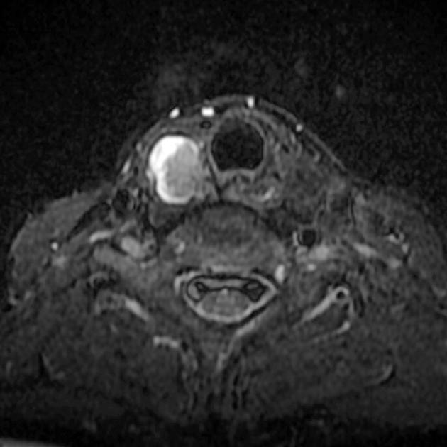
Denovo a complex thyroid nodule at the right lobe with a predominate intracystic solid nodule with mild hyperintensity (compared to muscles) on T1 and T2.
Case Discussion
50 year old patient with a previous papillary carcinoma of the thyroid on left treated with hemithyroidectomy, now presents with a new right sided nodule.
Patients with papillary thyroid cancer have an elevated risk for secondary primary cancer. Male gender and older age are recognized risks 1.
FNA
Smears reveal numerous cohesive fragments of follicular epithelium present mainly as loosely cohesive sheets attached to delicate branching capillaries or as densely cellular syncytial aggregates. Most of the follicular cells have ovoid, mildly pleomorphic irregular nuclei with fine chromatin and indistinct micronuclei. Many nuclei have nuclear grooves and a minor portion show nuclear inclusions. Some polygonal cells with dense "squamoid" cytoplasm are present. Colloid is scanty. A few fragments of follicular epithelium have follicular cells with scant cytoplasm, round nuclei and compact chromatin. This may represent sampling of adjacent benign epithelium. The features are sufficient for a diagnosis of papillary thyroid carcinoma.


 Unable to process the form. Check for errors and try again.