
Renal cell carcinoma extending to the renal vein and inferior vena cava
Diagnosis almost certain
Presentation
Gross hematuria.
Patient Data
Age: 65 years
Gender: Female


Download
Info

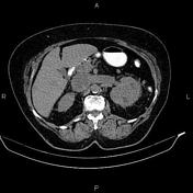
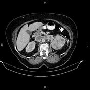
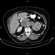
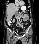
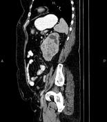
A 115×80×90 mm large hetero enhancing mass is noted at the left kidney that infiltrates the renal collecting system and extends to the left renal vein and IVC. Perinephric fat stranding is present. There is no sign of local invasion to other adjacent structures.
Several lymphadenopathies are observed in the vicinity of the mass with SAD less than 18 mm. A 45×30 mm lymphadenopathy is also noted at paraceliac region.
In imaged portions of the lower thorax, a few nodules are seen at lung fields less than 8 mm.
Case Discussion
Left renal mass; pathology proved renal cell carcinoma (clear cell subtype) with vascular extension, paraaortic and paraceliac lymphadenopathies, and lung metastases.


 Unable to process the form. Check for errors and try again.