
Presentation
Male with a history of 4 months of headache and seizures.
Patient Data
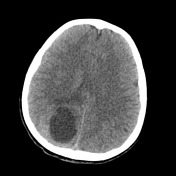

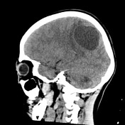

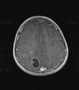
Homogenous hypodense well-circumscribed lesion in the periphery of the right parietal lobe associated with vasogenic perilesional edema.

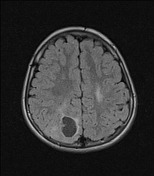
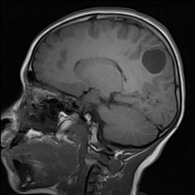

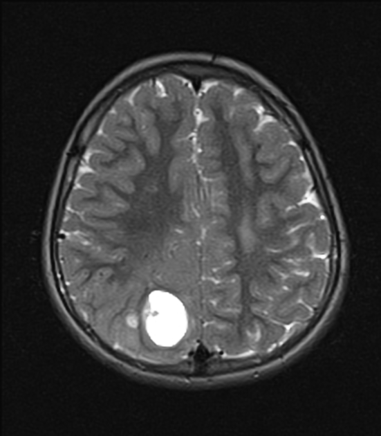
Well-circumscribed predominantly cystic lesion at the peripheral right parietal lobe ( precuneus) which is hyperintense in T2/FLAIR, hypointense in T1, after gadolinium administration with vivid ring and thick internal septa enhancement.
Case Discussion
Pleomorphic xanthoastrocytoma is a rare primary brain tumor that presents in young children and clinically manifests seizures and headaches, as in this case.
On MRI images appears as solid / cystic lesions located supratentorial and peripherally in cortex with leptomeningeal affection. Vividly enhancement of solid components and mural nodules is typical too. In this case, the images did not show wall nodules, however, it is observed a small amount of peripheral enhancement of solid tissue. After surgical resection pathology confirmed the presence of spindle cells, polygonal cells, multinucleated cells, and lipid infiltrated glial cells confirming the final diagnosis.


 Unable to process the form. Check for errors and try again.