
Presentation
Left flank pain and fullness.
Patient Data
Age: 75 years
Gender: Female
From the case:
Metastatic locally invasive renal cell carcinoma


Download
Info
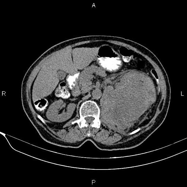
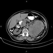
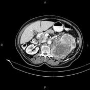
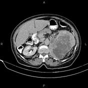
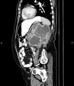
A 153×107×110 mm hetero enhancing mass is noted at the upper pole of the left kidney that invades adjacent psoas muscle and extends into the left renal vein and the proximal third of the left ureter.
A few lymphadenopathies are seen at para aortic regions.
A few small hypodense lesions are seen at the liver less than 10 mm that show no noticeable enhancement on delayed images, which could be suggestive of cysts.
A few tiny gallstones are observed.
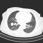
In imaged potions of lower thorax, multiple nodules are seen at both lungs less than 25 mm.
Case Discussion
The patient underwent left nephrectomy, and histopathology evaluation confirms renal cell carcinoma (clear cell type).


 Unable to process the form. Check for errors and try again.