
Presentation
Cough and dyspnea. Erythema nodosum of the lower extremities.
Patient Data
Age: 30 years
Gender: Male
From the case:
Sarcoidosis


Download
Info

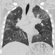
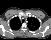
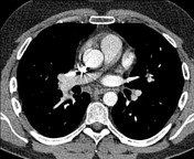
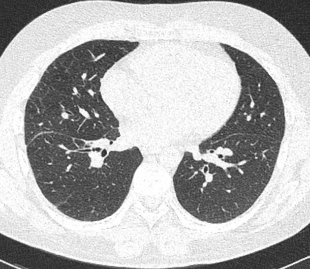
Mainly seen in the upper lobes:
- Many centrilobular nodularities, confluence of irregular margins, smaller nodules,
- Perilymphatic interlobular, intralobular interstitial and interlobar fissures thickening.
Enlarged bilateral hilar and mediastinal lymph nodes.
Histologically proven sarcoidosis.

Download
Info
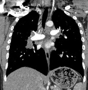
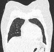
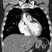
Compared with the previous CT, there is a noted regression of the disease as the affected areas have been decreased and the lymphatic nodes have been shortened. This means that the treatment for sarcoidosis has been effective.
Case Discussion
This is the II stage of sarcoidosis as both nodal and parenchymal involvement is present.
Nearly 90% of patients present with pulmonary manifestations. Patients between the ages of 20 and 40 are most often affected.


 Unable to process the form. Check for errors and try again.