Presentation
Cystic fibrosis patient. Suspicion of fatty infiltration of liver raised in report of abdominal ultrasound done 2 years earlier.
Patient Data
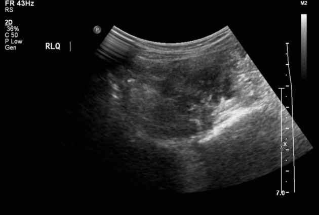
On scanning of the right lower abdomen, a mediocecal appendix is distended by thick secretions but its walls are not thickened, it exhibits hyperemia, and is incompressible. Minute amount of periappendiceal fluid. Importantly, no tenderness is elicited by pressure from the transducer.
The liver is of normal dimensions, exhibiting regular borders and moderate, diffuse, homogeneous hyperechogenicity.
The gallbladder is of normal dimensions, does not contain calculi, its walls are of normal thickenss.
The intra- and extrahepatic bile ducts are not distended.
The spleen is of normal dimensions, its echotexture is preserved.
The pancreas is masked by an abundance of air in the stomach.
The kidneys are of normal dimensions and structure. The parenchyma shows preserved thickness and echogenicity. Right kidney length is 7.9 cm, left kidney length is 8.5 cm. No evidence of collecting system dilatation.
The abdominal aorta shows normal diameter along its entire length.
The urinary bladder is moderately filled, the content is clear, its walls are normal.
In summary:
The liver shows moderate steatosis.
The appendix is distended by thick secretions, as can be expected given the underlying medical condition.
Case Discussion
The ultrasound examination revealed hepatic steatosis and an appendix distended by inspissated secretions in a child with an established diagnosis of cystic fibrosis.


 Unable to process the form. Check for errors and try again.