
Presentation
Work up for abdominal pain and gross hematuria.
Patient Data
Age: 50 years
Gender: Male
From the case:
Metastatic renal cell carcinoma


Download
Info
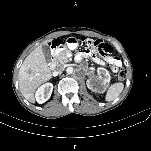
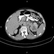
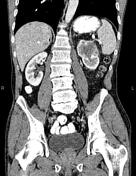
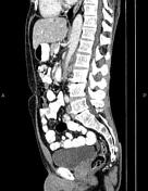
A 50×42 mm ill defined low enhancing mass is noted at the upper pole of the left kidney.
Several lymphadenopathy with a maximum SAD of 25 mm are noted at the left para-aortic regions. Most of them show internal necrotic changes.
Multiple low enhancing masses are seen at the liver less than 25 mm.
The prostate gland is enlarged.
Degenerative changes as osteophytosis are seen at the lumbar spine.
L5 vertebra is sacralized.
Case Discussion
Left kidney mass; pathology proved renal cell carcinoma (clear cell type) with para aortic lymphadenopathy and hepatic metastasis.


 Unable to process the form. Check for errors and try again.