
Presentation
Work up for hematuria.
Patient Data
Age: 55 years
Gender: Male
From the case:
Renal cell carcinoma


Download
Info
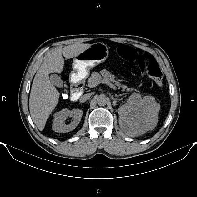
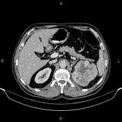
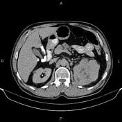
A 103×95×100 mm partially exophytic lobulated mass with heterogeneous enhancement and areas of necrosis is noted at the upper pole of the left kidney. Mild surrounding fat stranding is indicated, but no sign of local invasion to the adjacent structures, no vascular extension, and no regional lymphadenopathy.
Case Discussion
Left renal mass; pathology proved renal cell carcinoma (clear cell subtype).
On CT images, In general, small RCCs enhance homogeneously, whereas larger lesions have irregular enhancement due to areas of necrosis.
The clear cell subtype (70-80% of RCCs) is highly vascular and may show much stronger enhancement.


 Unable to process the form. Check for errors and try again.