
Presentation
Recent abdominal pain.
Patient Data


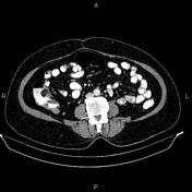
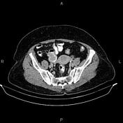
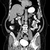
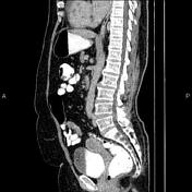
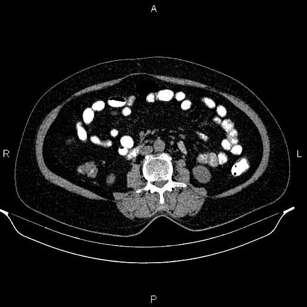
Short segment ileoileal intussusception is present at distal ileal loops, accompanied by mild bowel wall thickening and surrounding fat stranding. A 30×23 mm well-defined soft tissue density intraluminal mass is noted as the lead point.
Additionally, several enlarged mesenteric lymph nodes with SAD less than 18 mm are also observed at the RLQ.
Case Discussion
The patient underwent segmental ileal loop resection
Histopathology
Sections reveal intestinal tissue with the proliferation of spindle cells. Tumoral cells have faintly eosinophilic cytoplasm in a syncytial pattern. The overlying mucosa is unremarkable.
Immunohistochemistry evaluation was positive for DOG1, CD117 and CD 34 and negative for S100 and desmin.
Diagnosis: gastrointestinal stromal tumor (GIST)
Discussion
Although GIST can arise in any portion of the GI tract, from the esophagus to the rectum, the small bowel is the second most common site of involvement (20-25%) after the stomach (70%) and rarely may cause intussusception.


 Unable to process the form. Check for errors and try again.