
Presentation
Work up for abdominal pain and dyspepsia.
Patient Data


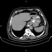
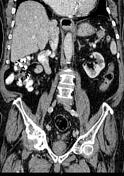
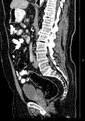
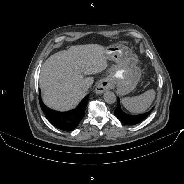
Increased wall thickness suggestive of tumoral infiltration is present at the esophagogastric junction, gastric cardia, subcardia, and proximal of the body, accompanied by perigastric fat stranding and several regional enlarged lymph nodes. Additionally, a few soft tissue density masses are observed in the upper abdomen suggest tumoral deposits.
Several non enhanced simple cortical cysts are seen in both kidneys.
The prostate gland is enlarged.
Degenerative changes as osteophytosis are seen at the lumbar spine.
Additionally, advanced degenerative changes are seen at the right hip joints.
Case Discussion
Pathology proved esophagogastric adenocarcinoma with regional enlarged lymph nodes and peritoneal seeding.


 Unable to process the form. Check for errors and try again.