
Presentation
Acute left lower quadrant pain.
Patient Data
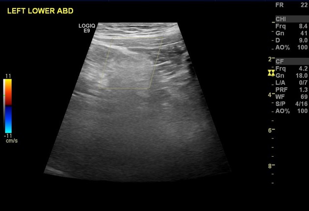
There is a hyperechoic mass-like abnormality in the left lower abdomen measuring 3 x 3 cm, non-compressible and without internal vascularity. It appears related to the bowel in the region likely due to epiploic appendagitis. CT scan is recommended for confirmation.
The rest of examination was unremarkable apart from diffuse fatty infiltration of the liver. (images are not included).
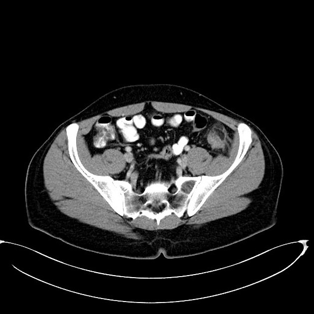
Fat density structure is seen anterior to sigmoid-descending colon junction with surrounding focal fat stranding. Associated with overlying peritoneal thickening surrounding the previously described structure "hyperattenuating ring sign "and minimal wall thickening of the adjacent colon, indicating epiploic appendagitis.
Fatty liver without focal lesion.
The appendix appears aerated measuring around 8 mm without surrounding inflammatory changes.
Case Discussion
This 25 year old man presented through ER with left lower quadrant pain. Along with initial labs abdomen and pelvis ultrasound was requested, showed hyperechoic mass at site of patient's complaint and guarding at this area. It appeared non-compressible without internal vascularity. No associated abdomen or pelvis free fluid. CT scan was advised by our radiology department and confirmed fat density lesion at typical location anterior to sigmoid-descending colon junction findings are those of epiploic appendagitis. Follow-up scan was also advised.


 Unable to process the form. Check for errors and try again.