
Patient Data
Note: This case has been tagged as "legacy" as it no longer meets image preparation and/or other case publication guidelines.
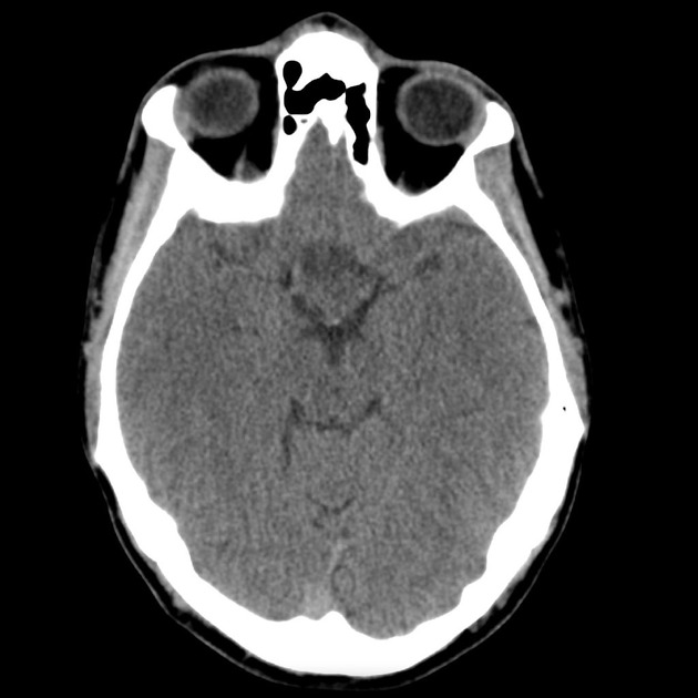
Suprasellar mass, predominantly of intermediate to fluid attenuation without convincing calcification.
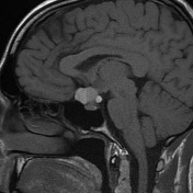
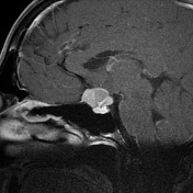
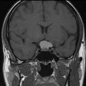
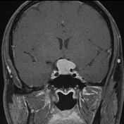

Selected sagittal and coronal pre and post contrast images demonstrate a multicystic/solid mass in the suprasellar region, with intrinsic high T1 signal.
Case Discussion
The patient went on to have a resection.
Histology:
The section shows a single small fragment of tissue. This is composed of irregularly shaped islands of mature squamous epithelium which are dispersed in a loose fibrous stroma. These show strong immunostaining for cytokeratins AE1/AE3 and CAM5.2. The stroma contains aggregates of keratin. Focal foreign-body type granulomatous inflammation in association with some collections of keratin are noted. There is also focal dystrophic calcification. No cyst formation is seen. The features are those of a craniopharyngioma.


 Unable to process the form. Check for errors and try again.