
Presentation
Subacute cognitive decline. Dizziness.
Patient Data


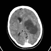

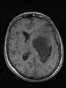
Hypodense left hemispheric lesion with mass effect. Midline shift, sulcal effacement and right lateral ventricle effacement. No enhancement with contrast.
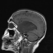
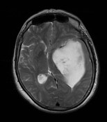
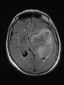
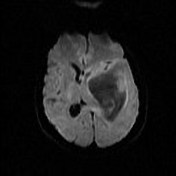

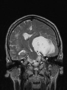
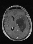
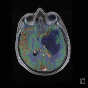

Left temporoinsular lesion with mass effect. High signal on T2 and FLAIR sequences. No diffusion restriction. In the lateral margin there is a focal area of enhancement with increased rCBV on perfusion weighted images.
Case Discussion
Stereotactic biopsy was performed. Histology confirmed the diagnosis of anasplastic astrocytoma (WHO grade III).
Note: IDH mutation status is not provided in this case and according to the current (2016) WHO classification of CNS tumors, this tumor would, therefore, be designated as an anaplastic astrocytoma NOS.


 Unable to process the form. Check for errors and try again.