
Presentation
Shortness of breath for investigation. GP had ordered a chest x-ray which demonstrated a mediastinal mass, which was further assessed with this non-contrast CT, performed at a community radiology practice. No contrast was administered due to the patient's borderline renal function.
Patient Data
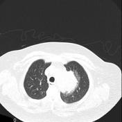

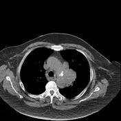

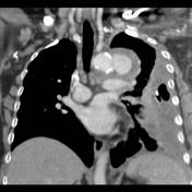
Non contrast CT demonstrates a mass abutting the arch of the aorta, without any intervening fat plane. The center of the mass is slightly hyperdense. A number of prominent lymph nodes are present in the mediastinum.
The provisional diagnosis of lung cancer, with likely direct invasion into the mediastinum (T4) was made.
Before further assessment the patient presented to the emergency department with increasing shortness of breath and left sided chest pain. This portable CXR was obtained.
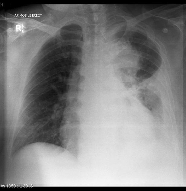
CXR again demonstrates the left sided mass, obscuring the outline of the aortic arch. A pleural effusion with associated atelectasis has developed.
A chest tube was inserted, yielding blood stained fluid.
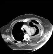

A planning CT is performed prior to transcutaneous biopsy of the mass. Due to the proximity of the mass to the aorta, despite the patients borderline renal function, a contrast bolus is administered. Contrast CT obtained for biopsy planning (see grid on anterior chest) demonstrates the mass to be a false aneurysm of the aorta rather than a pulmonary mass. Needless to say not biopsy was performed.
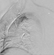
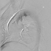
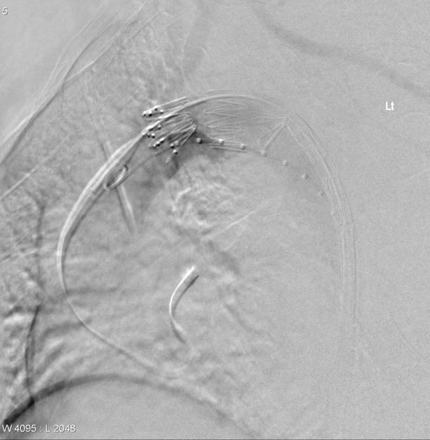
Endovascular aneurysm repair was performed and the patient had an uneventful recovery.
Case Discussion
This case illustrates the need for prudence when evaluating an anterior mediastinal mass, and accounts for one of the Ts in the familiar 5T mnemonic. The effusion was actually a hemothorax, and had a core biopsy of the mass been performed, it would no doubt have resulted in even further bleeding.


 Unable to process the form. Check for errors and try again.