
Patient Data
Age: Adult
Note: This case has been tagged as "legacy" as it no longer meets image preparation and/or other case publication guidelines.
From the case:
Arteriovenous malformation - cerebral
Show annotations
Download
Info
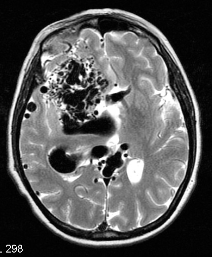
T2 axial imaging demonstrates numerous serpiginous flow-voids consistent with a very large right-sided arteriovenous malformation (AVM).
From the case:
Arteriovenous malformation - cerebral
Show annotations
Download
Info
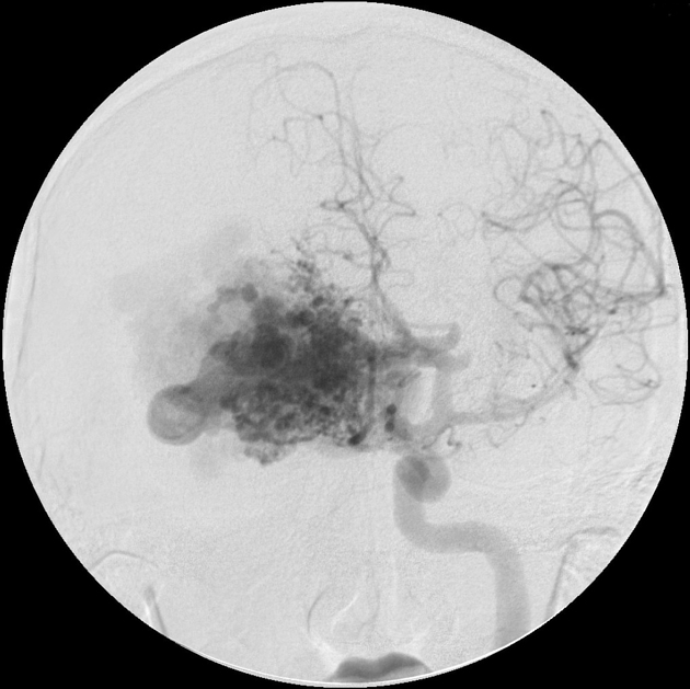
Angiography demonstrates arterial supply of a large right-sided nidus from both internal carotid arteries (only left ICA shown) and posterior circulation (not shown).
Case Discussion
Typical appearances of a large cerebral AVM.


 Unable to process the form. Check for errors and try again.