
Presentation
Leukocoria since birth, according to the parents.
Patient Data
Age: 1 year
Gender: Male
From the case:
Bilateral retinoblastomas

Show annotations
Download
Info

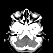
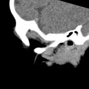
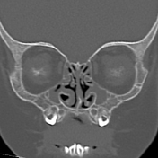
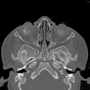
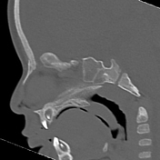
There are partially circumscribed hyperdense calcified intraocular lesions in retrolental regions on both sides; however, both eye globes are normal in size, shape, and position.
Additionally, opacification was incidentally noted in both the middle ear and mastoid.
Case Discussion
The CT features and the patient's age are highly indicative of retinoblastoma.


 Unable to process the form. Check for errors and try again.