
Presentation
Lower back pain
Patient Data
Age: 65 years
Gender: Female
From the case:
Breast cancer skeletal metastases

Download
Info
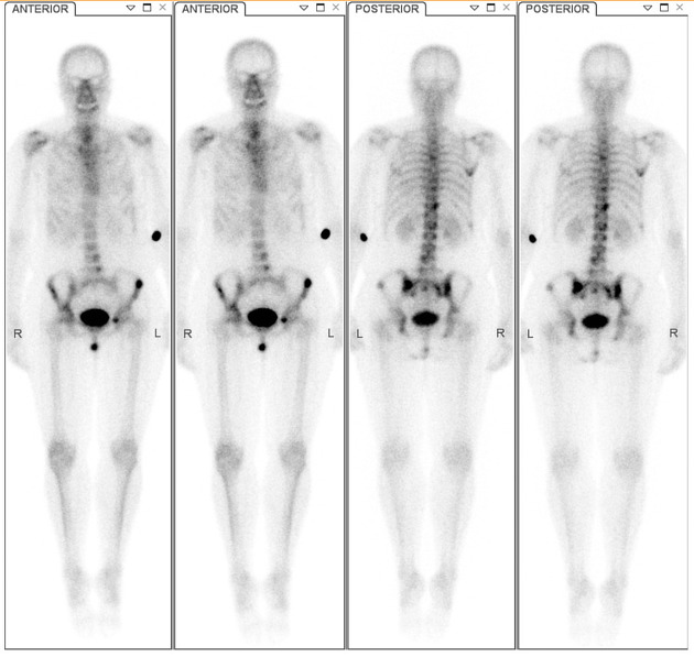
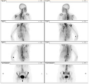
Delayed planar whole body planar and spot views demonstrate multiple foci of increased tracer accumulation in the axial and proximal appendicular skeleton. Lesions are most numerous in the pelvis, with the most intense lesions seen in bilateral posterior iliac bones and the left iliac crest.
Case Discussion
This is a case of osteoblastic metastatic disease in a patient with known breast cancer. Osteoblastic skeletal metastases demonstrate increased uptake of tracer (Tc99m HDP or MDP), reflecting the increased rate of matrix deposition associated with the lesions. On bone scan, skeletal metastases are more common in the axial and proximal appendicular skeleton.


 Unable to process the form. Check for errors and try again.