
Presentation
Altered mental status and headache of recent onset
Patient Data
Note: This case has been tagged as "legacy" as it no longer meets image preparation and/or other case publication guidelines.

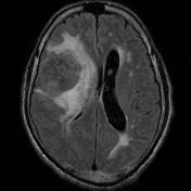

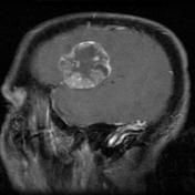

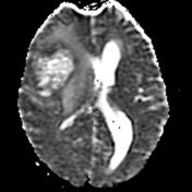
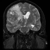
Large enhancing intra-axial mass of the right cerebrum with vasogenic edema, midline shift, subfalcine herniation and effacement of the ipsilateral lateral ventricle. Primary brain malignancy vs metastasis.
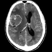
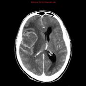
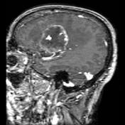


CT head: large enhancing intra-axial mass of the right cerebrum with vasogenic edema, midline shift, subfalcine herniation and effacement of the ipsilateral lateral ventricle. Primary brain malignancy vs metastasis.
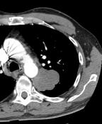

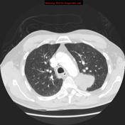
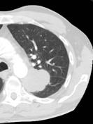

CTPA: lung mass centered on the superior segment of the left lower lobe, with invasion through the oblique fissure into the upper lobe, and also into the mediastinum. Centrilobular and paraseptal emphysema. Mass is suspicious for a primary bronchogenic carcinoma.
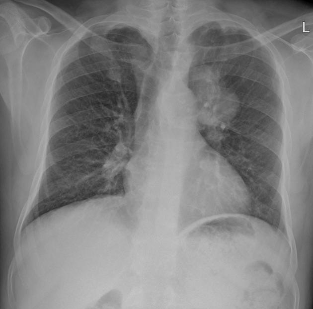
High suspicion for a bronchogenic carcinoma of the left upper zone.
Case Discussion
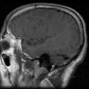
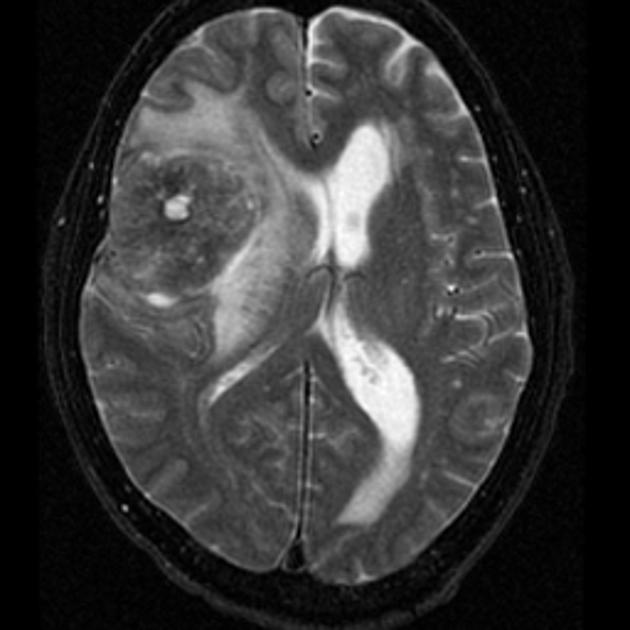
CT of the brain performed on-call revealed an enhancing intra-axial lesion. The possibility of a primary brain lesion was entertained. Given the patient's age, glioblastoma multiforme (GBM) was the primary consideration.
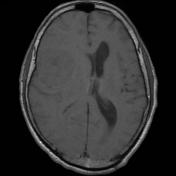
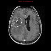
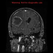
Brain MRI was performed the next day, and the differential diagnosis did not change, with GBM remaining high on the list.
The patient developed severe shortness of breath, and a CTPA was performed and revealed no pulmonary embolism, however, a large mass was noted. This was pathologically proven to be a bronchogenic carcinoma.


 Unable to process the form. Check for errors and try again.