
Central line in the accessory hemiazygos vein via the left superior intercostal vein
Diagnosis certain
Presentation
Post CVL insertion
Patient Data
Age: 27 week prem
Download
Info
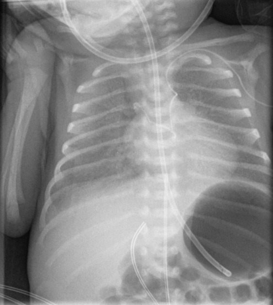
A small bolus of iodinated contrast was injected through the newly inserted left subclavian CVL at the time of X-ray acquisition. The CVL passes from the left brachiocephalic vein into the left superior intercostal vein and then the accessory hemiazygos vein. Injected contrast streams across the midline into the azygos vein and arch.
NGT in stomach. UVC in ductus venosus. Stable granular opacification in the lungs in keeping with known HMD. Resolving medial right PTX.
Case Discussion
Malposition of a CVL in a neonate. It was repositioned correctly.


 Unable to process the form. Check for errors and try again.