
Presentation
Knee pain after fall during football.
Patient Data
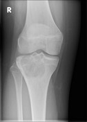
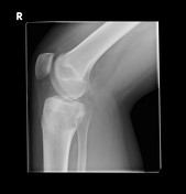

Large proximal tibial lucency centered on the metaphysis extending across the almost-fused physis into the epiphysis. Extension to the lateral tibial plateau joint surface. Possible central calcification. No periosteal reaction. No soft tissue mass.
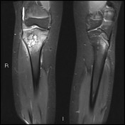

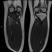
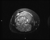
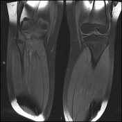
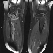
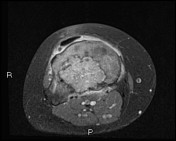

Right proximal tibial abnormality centered adjacent to the physis within the lateral metaphysis. Mixed solid and cystic components. Some fluid-fluid levels. Extension to the joint surface including the tibial spines. No pathological fracture.
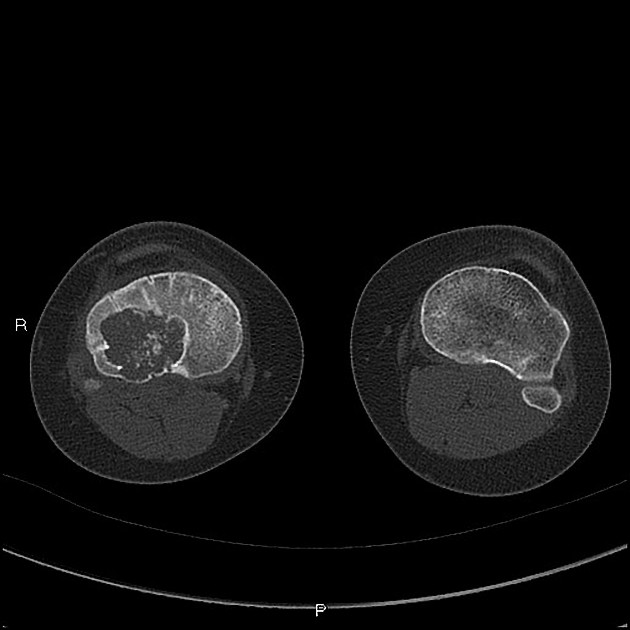
Well defined predominantly lucent tumor with central calcification (rings and arcs).
4 x 5 cm juxta articular lesion right tibial proximal metastasis. Thinning cortex proximally and posteriorly. Radiology appearances suggestive of giant cell tumor. Largely empty cavity with some lining curettage for specimen.
Right tibia, tissue from cystic cavity juxta-articular proximal tibia
Macroscopy
Multiple gritty fragments measuring up to 8 mm. All processed for decalcification.
Microscopy
Sections show multiple fragments of a bone tumor composed of plump oval histiocytoid cells with scatterd osteoclast-like giant cells. Within the cellular tissue there are islands of chondroid/cartilage. The appearances are consistent with those of a chondroblastoma.
Case Discussion
The plain film appearances are predominantly of a non-aggressive lesion. The MRI confirms its location and surrounding edema and the CT characterization of internal calcification is helpful. The differential, in this case, is of a chondroid lesion, with the most likely differential being chondroblastoma. This was confirmed by biopsy.


 Unable to process the form. Check for errors and try again.