
Presentation
Left foot pain.
Patient Data

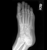

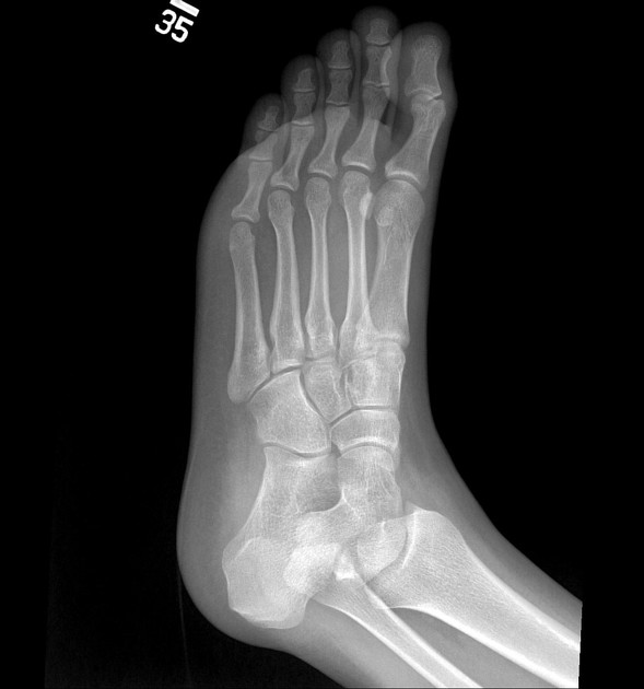
Subtle lucency and cortical ill-definition of the left intermediate cuneiform bone is noted as compared to the right intermediate cuneiform bone.


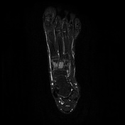
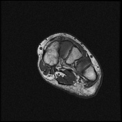
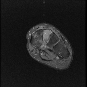
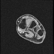
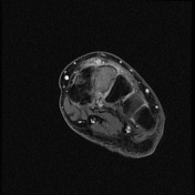
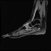
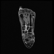
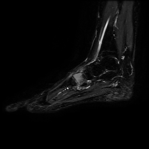
There is a lesion of isointense signal intensity to the muscle on T1 and of intermediate high signal intensity on T2 images occupying whole of the intermediate cuneiform bone and demonstrates enhancement post contrast administration. There are edematous changes of the adjacent cuneiform bones. Considering the patient's age and presentation, the most likely diagnosis is chondroblastoma.
Case Discussion
This case shows a rare presentation of chondroblastoma in the intermediate cuneiform bone. Chondroblastoma is one of the differential diagnosis of bone lesions that involve the epiphysis and the epiphyseal-equivalent region in a skeletally immature patient. The tumor is usually painful and on imaging classically demonstrates surrounding edema and enhancement. The patient underwent surgical resection and bone grafting. The diagnosis was confirmed by the histopathology report.


 Unable to process the form. Check for errors and try again.